Home

Naložený prísť maslo true lv aneurysm otto echo lev vybavenie Koncová tabuľka

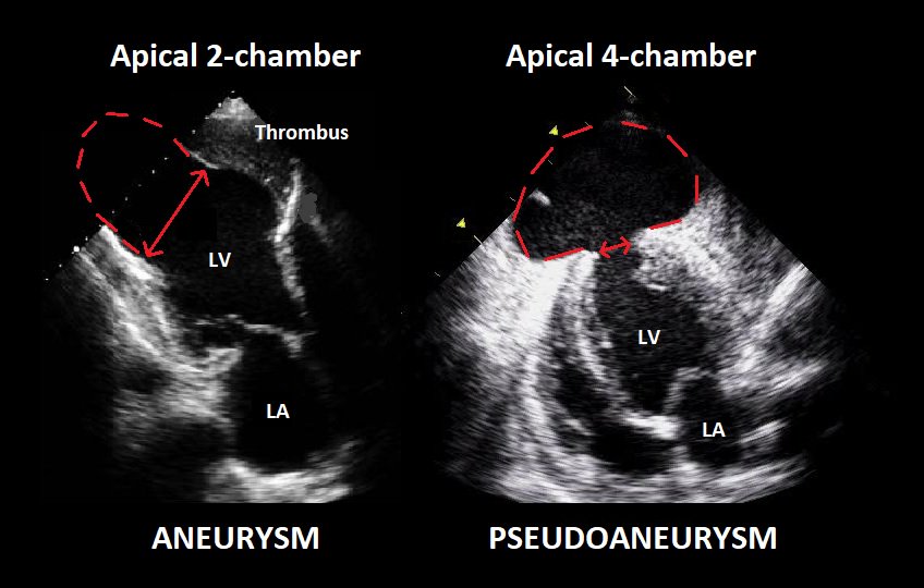
טוויטר \ Emergency Echo בטוויטר: "LV aneurysm: Wide neck, thinned  myocardial lining LV pseudoaneurysm: Narrow neck at myocardial rupture  site, pericardium/scar tissue lining https://t.co/9i4gMrycEI"
טוויטר \ Emergency Echo בטוויטר: "LV aneurysm: Wide neck, thinned myocardial lining LV pseudoaneurysm: Narrow neck at myocardial rupture site, pericardium/scar tissue lining https://t.co/9i4gMrycEI"
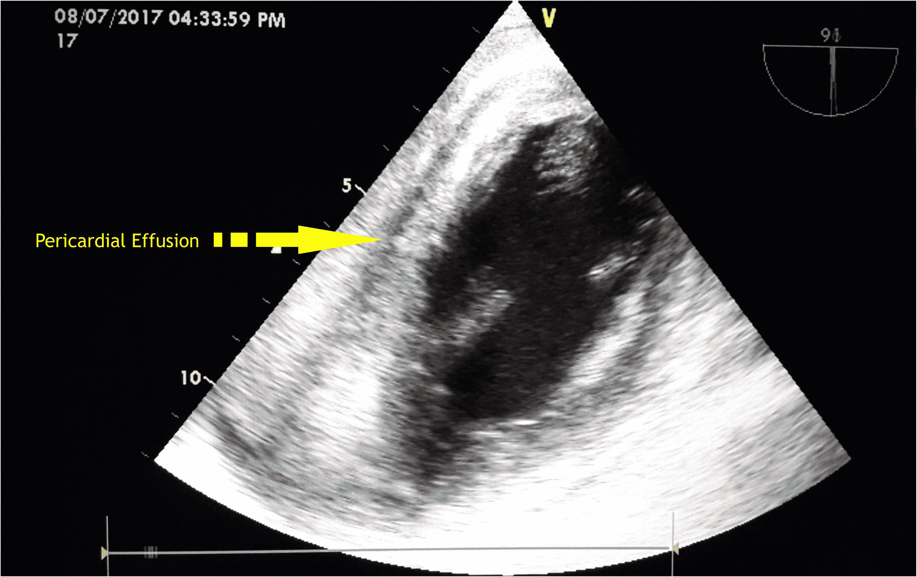
Cureus | Perioperative Management of a Patient With Left Ventricular Free  Wall Rupture After Myocardial Infarction: A Rare Case Scenario | Article
Cureus | Perioperative Management of a Patient With Left Ventricular Free Wall Rupture After Myocardial Infarction: A Rare Case Scenario | Article

A rare case of true and pseudoaneurysm of left ventricular wall and  incremental value of myocardial contrast Kalra GS, Tandon R, Singh B, Mohan  B - J Indian Acad Echocardiogr Cardiovasc Imaging
A rare case of true and pseudoaneurysm of left ventricular wall and incremental value of myocardial contrast Kalra GS, Tandon R, Singh B, Mohan B - J Indian Acad Echocardiogr Cardiovasc Imaging

Transesophageal echocardiography. Two-chamber view. LV pseudoaneurysm.... |  Download Scientific Diagram
Transesophageal echocardiography. Two-chamber view. LV pseudoaneurysm.... | Download Scientific Diagram

Transthoracic echocardiographic image of left ventricular septal... |  Download Scientific Diagram
Transthoracic echocardiographic image of left ventricular septal... | Download Scientific Diagram

Unruptured giant left ventricular pseudoaneurysm after silent myocardial  infarction | BMJ Case Reports
Unruptured giant left ventricular pseudoaneurysm after silent myocardial infarction | BMJ Case Reports
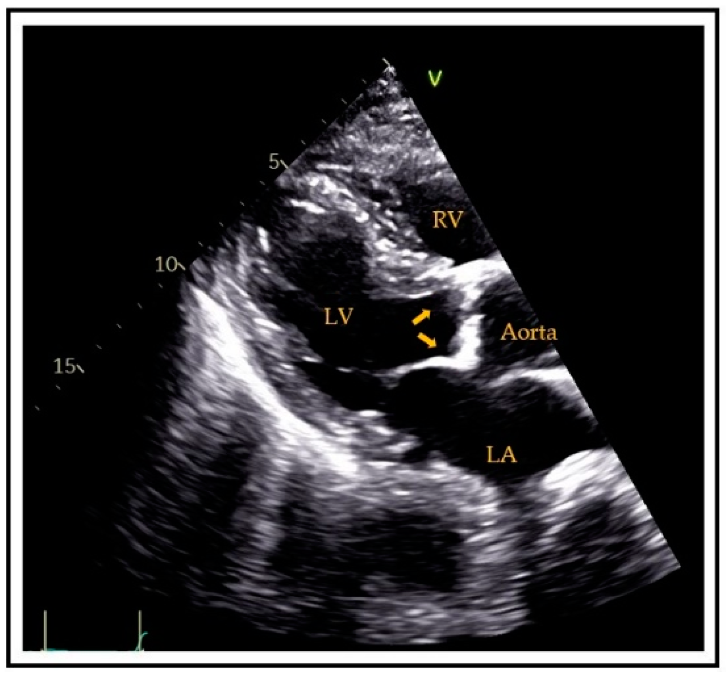
JCM | Free Full-Text | Cardiovascular Calcification as a Marker of  Increased Cardiovascular Risk and a Surrogate for Subclinical  Atherosclerosis: Role of Echocardiography
JCM | Free Full-Text | Cardiovascular Calcification as a Marker of Increased Cardiovascular Risk and a Surrogate for Subclinical Atherosclerosis: Role of Echocardiography

Anatomical and physiological complications related to left ventricular  apical aneurysm - Stoodley - 2017 - Sonography - Wiley Online Library
Anatomical and physiological complications related to left ventricular apical aneurysm - Stoodley - 2017 - Sonography - Wiley Online Library

Anatomical and physiological complications related to left ventricular  apical aneurysm - Stoodley - 2017 - Sonography - Wiley Online Library
Anatomical and physiological complications related to left ventricular apical aneurysm - Stoodley - 2017 - Sonography - Wiley Online Library

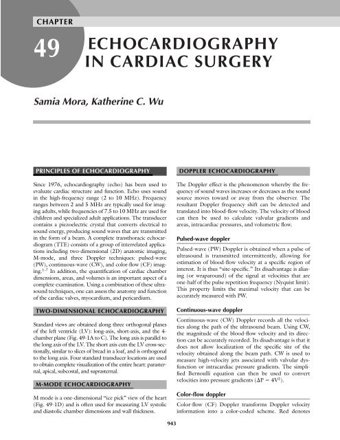
ECHOCARDIOGRAPHY IN CARDIAC SURGERY - TSDA

Echo-Web: Left Ventricular Aneurysm - YouTube

Echo assessment of coronary artery disease

A rare case of true and pseudoaneurysm of left ventricular wall and  incremental value of myocardial contrast Kalra GS, Tandon R, Singh B, Mohan  B - J Indian Acad Echocardiogr Cardiovasc Imaging
A rare case of true and pseudoaneurysm of left ventricular wall and incremental value of myocardial contrast Kalra GS, Tandon R, Singh B, Mohan B - J Indian Acad Echocardiogr Cardiovasc Imaging

Echocardiography - ScienceDirect

Giant apical LV aneurysm - 3D TTE

Left ventricular apical aneurysm associated with normal coronary arteries  following cardiac surgery: Echocardiographic features and differential  diagnosis - ScienceDirect
Left ventricular apical aneurysm associated with normal coronary arteries following cardiac surgery: Echocardiographic features and differential diagnosis - ScienceDirect

e 2-Dimensional Echo in apical 4 chamber view showing large LV apical... |  Download Scientific Diagram
e 2-Dimensional Echo in apical 4 chamber view showing large LV apical... | Download Scientific Diagram

Left Ventricular Apical Aneurysms in Hypertrophic Cardiomyopathy:  Equivalent Detection by Magnetic Resonance Imaging and Contrast  Echocardiography - Journal of the American Society of Echocardiography
Left Ventricular Apical Aneurysms in Hypertrophic Cardiomyopathy: Equivalent Detection by Magnetic Resonance Imaging and Contrast Echocardiography - Journal of the American Society of Echocardiography

Two-dimensional echocardiographic recognition of left ventricular  pseudoaneurysm.
Two-dimensional echocardiographic recognition of left ventricular pseudoaneurysm.

Echocardiography in the sub-costal view shows the left ventricular... |  Download Scientific Diagram
Echocardiography in the sub-costal view shows the left ventricular... | Download Scientific Diagram

Unruptured giant left ventricular pseudoaneurysm after silent myocardial  infarction | BMJ Case Reports
Unruptured giant left ventricular pseudoaneurysm after silent myocardial infarction | BMJ Case Reports
Medicowesome: LV aneurysm: Difference between true LV aneurysm and LV  pseudoaneurysm
Medicowesome: LV aneurysm: Difference between true LV aneurysm and LV pseudoaneurysm

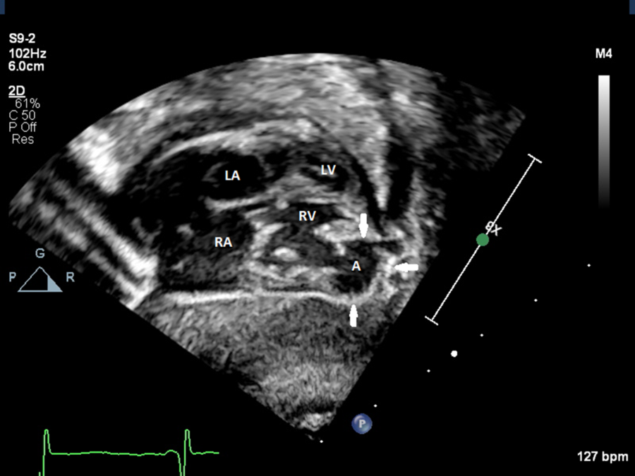
Congenital right ventricular aneurysm with characteristics of a  pseudoaneurysm | Cardiology in the Young | Cambridge Core
Congenital right ventricular aneurysm with characteristics of a pseudoaneurysm | Cardiology in the Young | Cambridge Core